When a baby is born without a fully formed ear canal, parents may hear several unfamiliar terms in rapid succession: congenital aural atresia, microtia, conductive hearing loss, canalplasty, bone-conduction hearing device, and ear reconstruction. It can feel as though the medical team has switched to a language composed entirely of complicated nouns.
The most important point is simpler: congenital aural atresia means the external ear canal did not develop normally. It may be completely closed or unusually narrow, and the eardrum and tiny middle-ear bones may also be underdeveloped. Some children have a normally shaped outer ear, but many have microtia, in which the visible ear is small, malformed, or absent.
Congenital atresia ear reconstruction can address hearing, appearance, or both. However, these goals are not always achieved through the same operation. Rebuilding the ear canal is different from constructing the visible outer ear, so successful care usually requires a coordinated team rather than one surgeon with an exceptionally ambitious afternoon.
What is congenital aural atresia?
The external auditory canal carries sound from the outside world to the eardrum. In congenital aural atresia, this canal is absent or blocked by bone and soft tissue. The condition may affect one ear, known as unilateral atresia, or both ears, known as bilateral atresia.
Although the ear canal and middle ear may not have developed normally, the cochlea and hearing nerve are often functional. Sound simply cannot travel through the usual pathway. This creates conductive hearing loss, meaning the problem occurs while sound is being conducted toward the inner ear rather than within the hearing nerve itself.
How atresia and microtia are connected
Aural atresia frequently occurs with microtia. Microtia is classified according to the extent of outer-ear development, ranging from a slightly small but recognizable ear to complete absence of the auricle, called anotia.
Microtia concerns the visible ear, while atresia concerns the ear canal. A child may therefore need two distinct forms of care:
- Hearing rehabilitation, such as a bone-conduction device or ear canal reconstruction.
- Auricular reconstruction to create a more typical-looking external ear.
Because treatment for one area can affect the timing or surgical approach used for the other, planning should involve an otologist or neurotologist, pediatric ear, nose, and throat specialist, audiologist, and reconstructive plastic surgeon.
What is the purpose of congenital atresia ear reconstruction?
The purpose depends on the operation being discussed. “Ear reconstruction” may describe functional reconstruction of the ear canal, cosmetic reconstruction of the auricle, or a coordinated combination of both.
Improving access to sound
Atresiaplasty, also called aural atresia repair or canalplasty, creates an open ear canal and reconstructs a pathway to the middle ear. When successful, it allows sound waves to reach the reconstructed eardrum and functioning middle-ear structures more efficiently.
Improved hearing may make it easier to understand speech in noisy classrooms, determine where sounds originate, and follow conversations when someone is speaking from the affected side. Children with atresia in both ears require especially early hearing support because limited sound access can interfere with speech and language development.
Creating a stable, usable ear canal
The surgical objective is not merely to drill an opening. The surgeon must create a canal that remains open, is lined with healthy skin, drains properly, and can be examined and cleaned over time. A poorly healed canal can narrow again, collect debris, or develop chronic drainage.
Reconstructing the visible ear
For children who also have microtia, outer-ear reconstruction can create an ear with a recognizable rim, bowl, folds, earlobe, and projection from the head. The reconstructed ear may make it easier to wear glasses or certain hearing devices and may reduce unwanted attention from other people.
Appearance is personal, however. Reconstruction is generally elective when performed solely to change the ear’s shape. Some children want surgery, while others prefer their natural ear or choose a prosthesis. The child’s opinion should become increasingly important as they grow.
How doctors evaluate a child for reconstruction
Not every child with aural atresia is a good candidate for canal surgery. Evaluation begins early, but final surgical planning often occurs several years later.
Newborn hearing screening and audiology testing
Babies should receive newborn hearing screening followed by diagnostic testing when an abnormality is found. Tests may include auditory brainstem response testing during infancy and behavioral hearing tests as the child becomes old enough to respond reliably to sounds.
Audiologists evaluate both ears, not only the visibly affected one. Even when atresia occurs on one side, the normally formed ear needs ongoing monitoring because it may become the child’s primary pathway for hearing.
Physical and developmental assessment
The medical team examines the outer ear, jaw, facial movement, palate, eyes, neck, and other craniofacial structures. Congenital ear differences can occur alone or as part of conditions such as hemifacial microsomia, Goldenhar syndrome, or Treacher Collins syndrome. A genetics consultation may be recommended when additional findings are present.
Temporal bone CT scan
A high-resolution computed tomography scan can show whether the middle ear, inner ear, facial nerve, and sound-conducting bones are developed well enough for canal reconstruction. Imaging is usually delayed until the child is closer to the age when surgery might be considered, commonly around age 5 or 6, unless an earlier medical concern requires investigation.
Surgeons may use the Jahrsdoerfer grading system to assess anatomical features on the scan. The score considers structures such as the stapes, middle-ear space, facial nerve position, and inner-ear anatomy. In general, a higher score suggests a better chance of useful postoperative hearing, but it does not guarantee a particular result.
Alternatives to ear canal reconstruction
Surgery is only one way to improve hearing. Many children use bone-conduction technology, either temporarily or as a long-term treatment.
Non-surgical bone-conduction devices
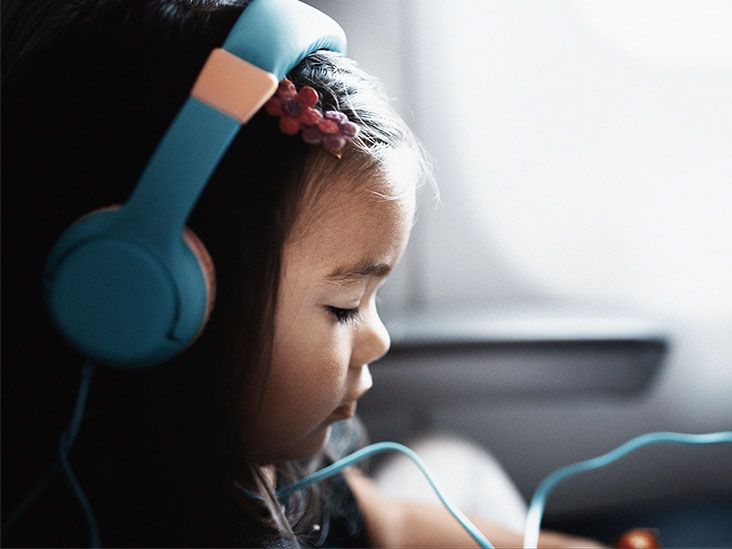
A bone-conduction processor sends sound vibrations through the skull directly to the functioning inner ear, bypassing the absent ear canal. Babies and young children may wear the processor on a soft headband, adhesive attachment, or another non-surgical support.
Early access to sound is particularly important when both ears are affected. Treatment should not be postponed for years simply because definitive reconstruction will occur later.
Implanted hearing systems
Older children may be candidates for an implanted bone-conduction system. Depending on the device, the processor connects through a small skin-penetrating abutment, a magnetic connection under the skin, or a fully implanted component that transmits vibrations to the bone.
These systems can provide predictable sound access without requiring the creation and lifelong maintenance of a new ear canal. Families should compare hearing quality, skin care, device visibility, surgical requirements, insurance coverage, and the child’s preferences.
How congenital aural atresia surgery is performed
Atresiaplasty is a delicate operation because the surgeon works close to the facial nerve, inner ear, jaw joint, and major blood vessels. The exact technique varies with the child’s anatomy.
Step 1: Coordinating the surgical sequence
If the child also wants microtia reconstruction, the ear canal surgeon and reconstructive surgeon decide which procedure should come first. The sequence depends partly on the type of outer-ear framework being used.
With rib-cartilage reconstruction, canal surgery is often performed after important stages of outer-ear reconstruction so incisions and blood supply are not disrupted. When a porous polyethylene framework is planned, atresia repair may be completed before or coordinated with auricular reconstruction. There is no universal schedule that fits every child.
Step 2: General anesthesia and surgical access
The child receives general anesthesia. The surgeon makes an incision near the malformed or absent canal and carefully exposes the temporal bone. Facial-nerve monitoring may be used because the nerve can follow an unusual path in children with congenital ear abnormalities.
Step 3: Creating the ear canal
Using a surgical drill, the surgeon removes bone to create a canal leading toward the middle-ear space. This process requires careful navigation around the facial nerve, jaw joint, inner ear, and any abnormally positioned structures.
Step 4: Reconstructing sound-conducting structures
The surgeon identifies the ossicles, the tiny bones that normally transfer vibration from the eardrum to the inner ear. If they are fused or malformed, they may be mobilized or reconstructed. In some cases, a prosthetic component is used to improve sound transmission.
A new eardrum is created with tissue such as fascia, a thin layer collected from beneath the scalp muscle. The graft is positioned so sound entering the new canal can vibrate the reconstructed membrane and middle-ear system.
Step 5: Lining and supporting the canal
The newly created bony canal must be covered with skin. Surgeons commonly use a thin split-thickness skin graft harvested from another area of the body, such as the scalp, thigh, or upper arm.
Packing material and a custom stent are placed inside the canal to support healing and reduce the risk of narrowing. Keeping the canal open is one of the most importantand sometimes most stubbornparts of recovery.
How the visible ear may be reconstructed
Outer-ear reconstruction does not directly restore hearing. Its purpose is to rebuild the auricle and improve symmetry.
Rib-cartilage reconstruction
In autologous reconstruction, the surgeon removes cartilage from the child’s ribs and sculpts it into an ear-shaped framework. The framework is inserted beneath the skin at the ear site. Later procedures may form the earlobe, elevate the ear away from the scalp, deepen the ear bowl, or refine individual contours.
This approach uses the child’s own tissue and can produce a durable reconstruction. It is usually delayed until the child has enough rib cartilage, often between ages 6 and 10, although timing varies among surgical programs.
Porous polyethylene reconstruction
A medical-grade porous polyethylene framework can be used instead of rib cartilage. The implant is covered with well-vascularized tissue, often taken from beneath the scalp, followed by a skin graft. This method may be offered at a younger age and may require fewer major stages, but implant exposure or infection can require revision.
Ear prosthesis
A realistic silicone prosthetic ear can be attached with medical adhesive or anchored to implants in the skull. A prosthesis may provide excellent visual detail without extensive cartilage reconstruction, although it requires daily care and periodic replacement as materials wear or the child grows.
Risks and possible complications
Congenital atresia reconstruction is complex, and families should receive a realistic discussion of benefits and limitations. Possible complications include:
- Re-narrowing or complete closure of the reconstructed canal.
- Chronic drainage, infection, or irritated tissue inside the canal.
- Skin-graft loss or delayed wound healing.
- Cholesteatoma, an abnormal collection of trapped skin cells.
- Damage to the facial nerve, which can weaken facial movement.
- Temporary or permanent changes in taste caused by nearby nerve injury.
- Inner-ear injury, dizziness, tinnitus, or worsening hearing.
- Scarring, asymmetry, framework exposure, or the need for revision surgery.
Restenosis, or narrowing of the reconstructed canal, is among the most common long-term concerns. Consistent stent use, cleaning, ear drops, and follow-up visits can help lower the risk but cannot eliminate it.
Recovery after atresia ear reconstruction
Recovery continues long after the child leaves the operating room. The ear may be covered with a bulky dressing, and packing usually remains in the canal until the surgeon removes or changes it.
Families receive instructions about pain medicine, antibiotic or steroid ear drops, bathing, sleeping position, and activity restrictions. The ear must generally remain dry during early healing. Swimming, contact sports, and rough play may be restricted until the surgical team confirms that the graft and canal are stable.
Once the initial packing is removed, a custom ear mold or stent may need to be worn for months. Follow-up appointments allow the surgeon to clean the canal, remove crusting, treat granulation tissue, and watch for narrowing. Audiograms are repeated to measure the functional hearing result.
A reconstructed canal also requires lifelong care. Some patients need periodic professional cleaning because the canal may not move wax and skin outward as efficiently as a naturally developed ear.
What results can families reasonably expect?
Well-selected patients can experience meaningful improvement in conductive hearing, but surgery does not guarantee normal hearing. Results depend on the original anatomy, movement of the middle-ear bones, healing of the eardrum and skin grafts, and whether the canal remains open.
A cosmetically reconstructed ear also will not be identical to a naturally developed ear. The goal is a balanced, recognizable ear that looks appropriate from ordinary conversational distancenot a microscopic duplicate suitable for fooling an ear anatomist at three inches.
Some families select canal surgery, while others choose a bone-conduction implant because it offers more predictable amplification and less canal maintenance. Others prioritize outer-ear reconstruction but continue using a hearing device. These are all legitimate choices when made with appropriate medical guidance.
Experiences families commonly have during the treatment journey
The first priority is usually hearing, not surgery
For many parents, the first months are filled with testing rather than reconstruction. The visible ear difference may be immediately apparent, but the medical team usually focuses first on determining how well each inner ear functions. Parents learn how to position a soft-band hearing device, keep it on a surprisingly talented little escape artist, and watch for responses to voices and everyday sounds.
Families often feel reassured when they understand that reconstruction does not need to be rushed. Hearing support, speech monitoring, and routine childhood development continue while the bones and cartilage grow. This creates time to research surgical options without treating every appointment as a countdown clock.
School can reveal challenges that are easy to miss at home
A child with normal hearing in one ear may speak clearly and respond well in a quiet room. Difficulties can become more noticeable in classrooms, cafeterias, playgrounds, or group activities. The child may turn the stronger ear toward a teacher, miss comments from one side, or have trouble identifying who called their name.
Parents commonly work with teachers on practical accommodations, such as preferential seating, reducing background noise, using a classroom microphone system, and checking that instructions were understood. These steps may appear small, but they can prevent a child from being mistakenly labeled inattentive when the real problem is acoustics.
The reconstruction decision can change over time
Some young children enthusiastically request a new ear. Others have little interest until they become more aware of appearance, glasses, hairstyles, sports equipment, or questions from peers. A child who wanted surgery at age 6 may feel differently at age 10, and the reverse can also happen.
Families frequently find it helpful to discuss all available choices without presenting one as the “correct” answer. Photographs, models, consultations with more than one surgical team, and conversations with people who have used different treatments can make the options less abstract. The goal is informed decision-making, not winning a family debate by producing the thickest binder.
Recovery requires organization and patience
The first days after surgery may involve swelling, tenderness, fatigue, dressings, and disrupted sleep. Children are often more interested in when they can return to sports than in the surgical masterpiece under the bandage. Parents, meanwhile, become temporary experts in medication schedules, dry-hair washing techniques, stent care, and detecting whether a sibling is approaching the healing ear at unsafe speed.
Follow-up care can be demanding, especially after canal reconstruction. Appointments for packing removal and canal cleaning may be uncomfortable. Stents can feel inconvenient, and ear drops may need to continue longer than expected. Families who live far from a specialty center should discuss local follow-up arrangements before surgery rather than discovering the travel burden afterward.
Emotional outcomes are individual
Some children feel more confident after reconstruction. Others were confident beforehand and view surgery mainly as a practical hearing decision. Children who do not choose cosmetic reconstruction may feel equally comfortable with their appearance, particularly when adults avoid treating the ear difference as something that must be hidden.
Questions from classmates are usually handled best with a brief, calm explanation the child can use comfortably: “I was born without an ear canal,” or “This device helps me hear.” Counseling, child-life services, school support, and contact with other families can help when teasing, anxiety, or self-consciousness becomes difficult.
The most positive experiences tend to occur when the treatment plan respects both medical needs and personal identity. The child is not merely an ear awaiting repair. They are a developing person whose preferences, hearing goals, daily routine, and comfort with surgery all matter.
Conclusion
Congenital atresia ear reconstruction can create a functional ear canal, reconstruct an underdeveloped outer ear, or coordinate both goals. Before surgery, specialists assess hearing, inner- and middle-ear anatomy, facial nerve position, developmental needs, and the child’s preferences.
Atresiaplasty creates a sound pathway by opening the temporal bone, reconstructing the eardrum and middle-ear mechanism, and lining the new canal with skin. Auricular reconstruction may use rib cartilage, a porous polyethylene framework, or a prosthetic ear. Bone-conduction devices remain an effective alternative or companion to reconstruction.
The best plan is individualized and should include realistic expectations about hearing improvement, appearance, recovery, canal maintenance, and possible revision procedures. Families should seek an experienced multidisciplinary program where hearing and reconstructive specialists plan treatment together.