Pneumonia already has a well-earned reputation for making people In some cases, however, the infection spreads beyond the lung and enters the narrow space between the lung and chest wall. Infected fluid or pus then collects in that space, causing a serious condition called empyema.
Empyema, also called empyema thoracis or pyothorax, can compress the affected lung, make breathing painful, and lead to sepsis or respiratory failure. It usually requires antibiotics and physical drainage of the infected fluid. Recognizing the causes, types, stages, and symptoms of empyema can help patients seek care before the infection becomes more difficult to treat.
What Is Empyema?
The lungs are covered by a thin membrane called the visceral pleura. Another layer, the parietal pleura, lines the inside of the chest wall. The small area between these layers is the pleural space. It normally contains a tiny amount of lubricating fluid that allows the lungs to glide smoothly while a person breathes.
Empyema occurs when microorganisms enter this normally sterile space and infected fluid or pus builds up. The collection can press against the lung and prevent it from expanding fully. Inflammation may also create strands of fibrin, separate pockets of fluid called loculations, and eventually a thick rind of scar-like tissue around the lung.
Empyema is related to a pleural effusion, but the terms are not interchangeable. A pleural effusion is any abnormal buildup of fluid around the lung. Heart failure, cancer, liver disease, blood clots, and pneumonia can all cause pleural effusions. Empyema specifically refers to an infected pleural collection, usually containing pus or other strong evidence of bacterial infection.
How Does Empyema Develop?
The most common pathway begins with bacterial pneumonia. Inflammation in the lung causes additional fluid to move into the pleural space. The fluid may initially be thin and free-flowing. If bacteria enter it, immune cells arrive, fibrin forms, and the fluid becomes increasingly cloudy, thick, and difficult to drain.
The Three Stages of Empyema
- Stage I: Exudative stage. Thin fluid collects around the lung because of nearby inflammation. The fluid may still be sterile, and doctors may describe it as an uncomplicated parapneumonic effusion rather than established empyema.
- Stage II: Fibrinopurulent stage. Bacteria and inflammatory cells enter the fluid. Pus and fibrin develop, and internal walls may divide the infection into separate pockets. The fluid is no longer behaving like water in a bowl; it is behaving more like pudding in a maze.
- Stage III: Organizing stage. Fibrous tissue forms a thick peel around the lung. This tissue can trap the lung and prevent normal expansion. Advanced disease may require surgery to remove the restrictive layer.
These stages describe a general progression, but patients do not always fit neatly into one category. Imaging, pleural-fluid findings, symptoms, oxygen levels, and the response to treatment all influence clinical decisions.
Common Causes of Empyema
1. Bacterial Pneumonia
Pneumonia is the leading cause of empyema. Bacteria can move from infected lung tissue into the pleural cavity, creating what is known as parapneumonic empyema. Organisms vary according to a patient’s age, underlying health, recent hospital exposure, previous antibiotics, and whether the pneumonia began in the community or a healthcare setting.
Frequently involved bacteria include streptococci, Staphylococcus aureus, and anaerobic bacteria that normally live in the mouth. More than one organism may be present. Pleural-fluid cultures can also be negative, especially when antibiotics were started before fluid was collected.
2. Lung Abscess and Aspiration
A lung abscess is a pocket of infected, damaged tissue inside the lung. If the infection spreads outward or the abscess ruptures, bacteria may enter the pleural space.
Aspiration can produce a similar problem. Food, saliva, or stomach contents may enter the lungs when normal swallowing or coughing reflexes are impaired. Aspiration risk can increase with seizures, neurologic disorders, severe swallowing difficulties, reduced consciousness, or heavy alcohol use.
Poor oral hygiene is another risk factor because bacteria from the mouth may be carried into the lower respiratory tract. Dental care may not sound like lung care, but the mouth and lungs are surprisingly close business partners.
3. Chest Surgery or Trauma
Empyema may develop after lung surgery, esophageal surgery, chest-tube placement, or another procedure involving the chest. Penetrating injuries, blunt trauma, and an infected collection of blood around the lung can also introduce bacteria into the pleural cavity.
4. Esophageal Rupture
A tear in the esophagus can release bacteria-rich material into the chest. This may cause a severe pleural and mediastinal infection that requires emergency treatment. Infection can also spread from an abscess beneath the diaphragm, infected ribs, the spine, or other nearby structures.
5. Tuberculosis and Fungal Infection
Tuberculosis can cause chronic pleural infection and empyema, particularly when diagnosis or treatment is delayed. Fungal empyema is uncommon and is more likely in people with significant immune suppression, prolonged hospitalization, recent surgery, or an esophageal leak.
Types of Empyema
Doctors may classify empyema according to its cause, duration, anatomy, or appearance on imaging.
Parapneumonic Empyema
This is the most common type. It develops as a complication of pneumonia and represents the infected end of the parapneumonic-effusion spectrum.
Postoperative Empyema
Postoperative empyema occurs after chest surgery. Postpneumonectomy empyema, which develops after removal of a lung, is a particularly complex form because the normal anatomy of the chest has been altered.
Post-Traumatic Empyema
This type follows a chest injury, an infected hemothorax, or contamination of the pleural space after trauma.
Acute Empyema
Acute empyema develops over days or weeks. It commonly causes fever, pleuritic chest pain, cough, rapid breathing, and systemic illness.
Chronic or Organizing Empyema
Chronic empyema persists long enough for dense pleural thickening and scarring to develop. The affected lung may become trapped, leading to ongoing breathlessness, fatigue, weight loss, and reduced exercise tolerance.
Loculated Empyema
A loculated empyema contains several separated pockets rather than one freely moving collection. Loculations can make treatment more complicated because a drainage tube positioned in one pocket may not empty the others.
Empyema Necessitans
Empyema necessitans is a rare complication in which infection extends through the pleura and into the chest-wall tissues. It may appear as a painful swelling, mass, or draining sinus and requires urgent specialist treatment.
Empyema Symptoms
Empyema symptoms often resemble pneumonia. One of the most useful warning signs is a recovery that stops progressing. Fever may return, breathing may become more difficult, or chest pain may persist despite antibiotics.
Common Symptoms in Adults
- Fever and chills
- Night sweats
- Shortness of breath
- Rapid, shallow breathing
- Sharp chest pain that worsens with coughing or deep breathing
- Dry or productive cough
- Extreme tiredness or weakness
- Loss of appetite
- Unintentional weight loss
- Fast heart rate
- Pneumonia symptoms that persist or worsen during treatment
Older adults and people with weakened immune systems may not develop an obvious fever. Confusion, poor appetite, unusual sleepiness, falls, or sudden loss of independence may be more noticeable than classic respiratory symptoms.
Symptoms in Children
Children may experience fever, cough, rapid breathing, chest or abdominal pain, poor feeding, lethargy, and irritability. A child with pneumonia who remains feverish, increasingly breathless, or unusually exhausted after treatment begins should be reassessed.
Emergency Warning Signs
Emergency care is necessary for severe breathing difficulty, bluish or gray lips, confusion, fainting, inability to stay awake, very low oxygen levels, rapidly worsening chest pain, or signs of sepsis such as clammy skin, extreme weakness, fast breathing, and a racing heart.
Who Is at Greater Risk?
Anyone can develop empyema, including previously healthy children and adults. Risk may be higher among people with diabetes, bronchiectasis, chronic lung disease, cardiovascular disease, liver disease, cancer, immune suppression, poor nutrition, aspiration disorders, harmful alcohol use, intravenous drug use, or poor oral health.
Recent hospitalization, chest surgery, mechanical ventilation, and repeated pneumonia can also increase risk. Older adults may be especially vulnerable to serious complications, although empyema can occur at any age. Unfortunately, the pleural space does not check birth certificates before becoming infected.
How Empyema Is Diagnosed
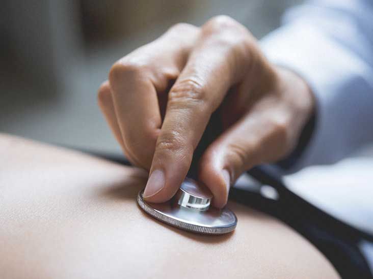
Diagnosis begins with a medical history, physical examination, oxygen measurement, and review of pneumonia symptoms. A clinician may hear reduced breath sounds or a rubbing sound over the affected area.
Chest Imaging
A chest X-ray may show fluid around the lung. Ultrasound can identify small collections, reveal internal septations, and guide safe placement of a needle or drainage tube. Contrast-enhanced CT provides more detailed information about pleural thickening, loculations, lung abscesses, and other chest abnormalities.
Pleural-Fluid Testing
During thoracentesis, a clinician removes pleural fluid with a needle. The sample may be evaluated for appearance, acidity, glucose, enzymes, white blood cells, Gram stain, and bacterial culture. Frank pus strongly indicates empyema. A negative culture does not completely exclude infection.
Blood Tests
Blood tests help evaluate inflammation, organ function, and overall severity. Blood cultures may detect bacteria circulating in the bloodstream. No single result tells the entire story, so clinicians interpret the findings together rather than holding a laboratory talent contest.
How Is Empyema Treated?
Empyema generally requires hospital treatment. The two main goals are to control the infection and remove the infected collection so the lung can expand.
Antibiotics
Doctors begin antibiotics that cover likely organisms and adjust the medication when culture results or other clinical information becomes available. Treatment may continue for several weeks, depending on the cause, severity, drainage success, and response.
Chest-Tube Drainage
Infected or purulent fluid usually needs to be drained through a chest tube. Imaging guidance helps position the tube in the collection. It may remain in place for several days while doctors monitor drainage, fever, breathing, laboratory results, and repeat scans.
Intrapleural Medicines
Some patients with loculated infection receive medicines through the chest tube to break down fibrin and make thick material easier to drain. A combination of tissue plasminogen activator and deoxyribonuclease may be considered, although it is not appropriate for every patient.
Surgery
Video-assisted thoracoscopic surgery may be used to wash out infected material and open loculated pockets. Decortication removes the thick fibrous peel surrounding a trapped lung. Open surgery is sometimes necessary when minimally invasive treatment is unsuccessful or the infection is advanced.
Complications and Outlook
Possible complications include sepsis, respiratory failure, pleural scarring, trapped lung, reduced lung function, recurrent infection, bronchopleural fistula, and extension of infection into the chest wall.
Many patients recover well when the condition is recognized and treated promptly. Recovery depends on the underlying cause, stage of infection, general health, and success of drainage. Fatigue and reduced stamina may continue after discharge, and follow-up imaging may be needed to confirm that the fluid has resolved.
Can Empyema Be Prevented?
- Seek care when pneumonia symptoms are severe, persistent, or worsening.
- Take prescribed antibiotics exactly as directed.
- Return for evaluation if fever or breathlessness continues during treatment.
- Follow current influenza and pneumococcal vaccination recommendations.
- Maintain good oral and dental hygiene.
- Address swallowing difficulties and aspiration risks.
- Follow wound-care instructions after chest surgery.
- Avoid smoking and seek support for harmful alcohol or drug use.
Not every case can be prevented, but prompt treatment of pneumonia and other lung infections may stop pleural fluid from progressing to empyema.
Experiences Related to Empyema: What the Journey May Feel Like
The following are composite educational scenarios based on common patterns of care. They are not quotations from individual patients and should not be used for self-diagnosis.
When Pneumonia Recovery Stalls
A common experience begins with fever, cough, chest soreness, and exhaustion. Antibiotics are prescribed for pneumonia, and everyone expects a gradual turnaround. Instead, several days pass and the fever keeps returning. Walking from the bedroom to the bathroom feels like an endurance event. A sharp pain appears with every deep breath, so the person unconsciously begins breathing more shallowly.
It can be easy to rationalize these changes. Pneumonia is tiring, coughing hurts, and recovery does take time. The important clue is the direction of travel. When breathing becomes harder, pain intensifies, or energy continues to decline rather than gradually return, another examination is needed. Repeat imaging may reveal a pleural collection that was previously too small to detect.
The Hospital and Chest-Tube Experience
Many patients are surprised to learn that antibiotics alone may not solve the problem. The infection is not only inside the lung; infected material is trapped outside it. Doctors may use ultrasound or CT to plan drainage before placing a chest tube between the ribs.
The tube may feel sore, awkward, or as though it pulls during movement. Nurses monitor the drainage system and help the patient change positions safely. Effective pain control is important because comfortable breathing and coughing help the lung expand.
Patients may use an incentive spirometer, sit in a chair, walk with assistance, and work with respiratory or physical therapists. Progress is not always linear. One morning may bring more energy, while the afternoon brings a nap long enough to qualify as a temporary relocation.
When the First Drain Is Not Enough
Loculated empyema can be frustrating because one drainage tube may not reach every pocket. Follow-up imaging may show that part of the collection remains. The medical team might reposition the tube, place another drain, administer intrapleural medicines, or recommend thoracoscopic surgery.
Hearing the word “surgery” after spending days in the hospital can feel as though the illness has added an unnecessary sequel. However, opening loculations and removing restrictive tissue may finally allow the lung to expand and the fever to resolve.
Understanding the purpose of each treatment can make the process less frightening. Antibiotics fight the organisms, drainage removes infected material, and surgery addresses pockets or scar tissue that a tube cannot fully clear.
Recovery After Discharge
Going home does not necessarily mean feeling normal. Stamina may remain limited, appetite may return slowly, and the chest-tube or surgical site can stay tender. Someone who normally climbs stairs without thinking may need several pauses.
Helpful recovery habits include completing antibiotics, attending follow-up visits, increasing activity gradually, eating enough protein and calories, staying hydrated, and following instructions for incision care. New fever, worsening shortness of breath, increasing chest pain, confusion, drainage from an incision, or a sudden decline should prompt immediate medical advice.
The Emotional Side of Empyema
Empyema can be emotionally exhausting as well as physically painful. An unexpected hospitalization may disrupt work, caregiving, finances, and sleep. Some people become anxious about every cough after recovery. Others blame themselves for not seeking help earlier, even though the first symptoms often resemble an ordinary respiratory infection.
A practical recovery plan can reduce some uncertainty. Patients may write down warning signs, carry an updated medication list, arrange help at home, and track gradual improvements in walking, appetite, sleep, and breathing.
Recovery is not a competition. The lungs do not award medals for pretending everything is fine, and asking for help is considerably more useful than attempting to win an imaginary toughness championship.
Conclusion
Empyema is a serious infection in which pus or infected fluid collects in the pleural space, most often after pneumonia. The disease may progress from thin fluid to loculated pus and eventually to a fibrous peel that traps the lung.
Fever, cough, fatigue, pleuritic chest pain, and shortness of breath are common symptoms. A pneumonia infection that fails to improve or suddenly worsens is an especially important warning sign. Diagnosis usually involves chest imaging and pleural-fluid testing, while treatment generally combines antibiotics with drainage. In complex cases, intrapleural medicines or surgery may be needed.
Early evaluation matters because untreated empyema can lead to sepsis, respiratory failure, and permanent pleural scarring. Seeking medical attention is not overreacting; it is giving the infection less time to wrap the lung in the world’s least welcome blanket.